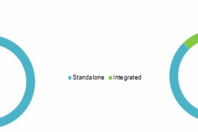

Cone Beam Computed Tomography (CBCT) is a cutting-edge imaging technique with numerous clinical uses in dentistry. Cone-beam computed tomography has expanded in use throughout time and is primarily thought to be helpful in the diagnosis and planning of therapy for implant dentistry, endodontics, ENT, maxillofacial surgery, and other procedures. Integrated CBCT is also used to position patients during operations. Because modern computers outfitted with CBCT units are becoming increasingly affordable, the use of cone-beam computed tomography (CBCT) systems has increased dramatically.
Numerous dentists and imaging specialists frequently use and even recommend these methods to pinpoint the issue with absolute precision. A variant of conventional computed tomography (CT) systems are Cone Beam Computed Tomography (CBCT) systems. Dental professionals use CBCT machines, which revolve around the patient and use a cone-shaped X-ray beam to collect data. Cone Beam CT gathers the required information in a single cycle using a bigger imaging area than normal CT and rotating the subject 360 degrees.
Read More @ https://cmibloginsight.blogspot.com/2022/08/cone-beam-computed-tomography-provides.html