Excision of Fibroma using Dental Soft-tissue Diode Laser
Introduction- A fibroma is a benign scar like reaction due to persistent long standing irritation in the mouth resulting in an increase in connective tissue at the site of repeated trauma. They are also known as traumatic fibromas, focal intra-oral fibrous hyperplasia, fibrous nodules, or oral polyps.
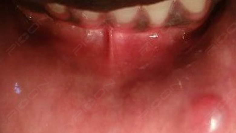
Clinical Features- Fibromas are often found on the interior of the cheek, lips, or lateral borders of the tongue and result from tissue being trapped between the maxillary and mandibular teeth when occluding. An oral fibroma presents as a firm smooth lump and is usually similar in color to the surrounding soft tissue, although it may be paler due to less vascularity and increased connective tissue within it. A fibroma also may present darker if trauma has led to bleeding inside of it and may have an ulcerated surface related to recent trauma. Fibromas have a dome-shaped geometry but may be pedunculated (polyp-like with a short stalk-like base).
Treatment Biopsy and oral fibroma removal traditionally has been performed with a scalpel that results in excision resulting in bleeding margins. The raw bleeding edge left by scalpel excision frequently requires suture placement to control bleeding. Additionally, these types of wounds have been associated with postoperative pain and inflammation. The use of sutures for wound management may exacerbate site irritation until their resorption or removal. Later techniques advocated the use of monopolar electrosurgery as an alternative to the scalpel blade. Electrosurgery, if used improperly, has the potential to cause tissue burns and increased site inflammation that may hamper healing.
With the advent of lasers for soft-tissue surgeries, various types of laser devices have been used successfully for oral fibroma removal. Laser selection is practitioner dependent. PIOON Laser offers different wavelengths like 450nm/810nm or 980nm. The most preferred among them is 450nm wavelength. After informed consent, the lesion was infiltrated with local anesthesia, the protective eye wear were worn and then the lesion was excised using 450nm in noncontact mode with surgical tip (400µm) making sure to remove the stalk attaching the lesion. After the procedure, postoperative instructions were given and analgesic was prescribed on as and when basis.
Also, care should be taken to get rid off the cause (sharp cusps or fractured tooth if any) along with the excision to make the treatment outcome more predictive. Here low level laser therapy can also be done over the exposed wound area using 660nm (red light) in noncontact mode to assist in faster healing and to manage postoperative pain.
Rationale of use of lasers - An advantage of diode laser application compared to scalpel use is the provision of a relatively bloodless surgical incision with minimal swelling and scarring postoperatively. Also, compared to an electrosurgery tip, the use of a diode laser reduces the zone of affected cells considerably, thus less tissue trauma and thermal burning and better healing have been reported. Consequently, the diode laser is suitable for excision of oral fibromas.
An interesting study by C. Fornaini et al suggested decreased excision time with a blue diode laser (wavelength 450nm). The operating time was considerably decreased and the superficial rise in temperature at the operated site was markedly lower as compared to that with 810 or 1370 nm wavelength laser.
Conclusion- Dental Diode lasers can be used for fibroma removal. A 450 nm wavelength offers significantly better healing with comfort for the patient themselves.
References - Carlo Fornaini, Jean-Paul Rocca, Elisabetta Merigo. 450nm blue laser and oral surgery: preliminary ex vivo study. The Journal of Contemporary Dental Practice, October 2016;17(10):795-800