views

Management of Pericoronitis using Dental Diode Laser
Introduction- The operculum is a soft tissue covering the crown of a partially erupted tooth. This area is difficult to access, and hence the normal oral hygiene methods remain ineffective, leading to inflammation of the soft tissue. This inflammation of the soft tissue surrounding the crown of a partially erupted tooth is called pericoronitis. It is known to appear when the third molars start to erupt, and are most commonly seen with the partially erupted/impacted mandibular third molars. Pericoronitis is treated by extraction of the third molar or by performing operculectomy. Operculectomy is a procedure which is done by excising the excessive soft tissue covering the third molar. It is indicated when there is availability of space for eruption of third molar, presence and proper alignment of antagonist tooth and impacted third molar in the arch.
Etiology- The most common cause behind pericoronitis is the entrapment of plaque and food debris between crown of tooth and overlying gingival flap or operculum and also the constant trauma of opposing maxillary third molar causes pericoronal inflammation.
Treatment modality -The painful or inflamed/ infected operculum can be removed by a wide variety of techniques like scalpel, caustic agents, radiofrequency surgery, electrocautery etc. The drawbacks of these techniques are intraoperative and post-operative bleeding, need for anaesthesia, suturing, risk of post-operative infection, edema and impaired visibility due to bleeding.
However, Laser therapy is an effective and noninvasive treatment option for Operculectomy. PIOON Laser offers different wavelengths like 450nm/810nm or 980nm. Out of which 450nm can be used in non-contact/contact mode whereas 810nm and 980nm are used in contact mode to perform operculectomy.
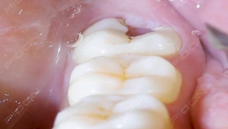
In this case, on the basis of radiographic and clinical findings, diagnosis of pericoronitis was made and operculectomy procedure was planned. Wavelength of 450nm of PIOON laser was used in noncontact mode for the excision of inflamed pericoronal flap under topical anesthesia and crown of the carious tooth was exposed which further cleared the path of eruption. Post-operative instruction along with maintenance of oral hygiene was advised. The complete safety protocols were followed for the patient, operating and assistant staff like using laser protective eye glasses and use of high vacuum suction. Highly reflective instruments were avoided while using lasers.
Khan MA et al in 2017 used 980nm wavelength for operculectomy and suggested that due to many intraoperative and postoperative advantages, diode laser has become a preferred option for minor oral surgical procedures. Fornaini C et al. in 2016 also concluded that the 450 nm wavelength works very efficiently in the oral soft tissue surgical procedures, with faster healing and lesser thermal damage.
Rationale behind Use of Lasers- Lasers offer certain advantages like the excellent hemostatic property which is of great value and allows the surgeon to work with better visibility, minimum patient discomfort, reduced post-operative pain, less chair side time, reduced healing time, reduction in edema and post-operative bleeding.
Conclusion – Taking into consideration the excellent clinical outcome, the dental diode lasers can be considered as a better and acceptable alternative for excision of pericoronal flap.
References - Khan MA et al. Diode laser: A novel approach for the treatment of pericoronitis. J Dent Lasers 2017;11:19-21 and Fornaini C et al. 450 nm diode laser: a new help in oral surgery. World J Clin Cases 2016; 4:253–257.

Preoperative View (Courtesy- Dr. Sana Farista)

Intraoperative view (Courtesy- Dr. Sana Farista)

Postoperative View (Courtesy- Dr.Sana Farista)


