FRENOTOMY USING DIODE LASER
Frenum is an anatomic structure formed by a fold of mucous membrane and connective tissue fibers that attach the lip and cheeks to the alveolar mucosa, the gingiva and the underlying periosteum. Placek et al. have classified frenum depending on the extension of attachment of fibers, (1) Mucosal: when the frenal fibers are attached up to mucogingival junction, (2) Gingival: when fibers are inserted within attached gingiva, (3) Papillary: when fibers are extending into interdental papilla, and (4) Papilla penetrating: when the frenal fibers cross the alveolar process and extend up to palatine papilla.
Frenum with mucosal attachment is the usual and is considered to be the correct level of attachment but if the frenum that encroaches on the margin of the gingiva (Class 2 and above) may interfere with plaque removal, and the tension on the frenum may tend to open the sulcus eventually leading to gingival recession, midline diastema, root dentine hypersensitivity and unaesthetic looks. Nowadays aesthetic concerns have led to an increasing importance in seeking dental treatment, with the purpose of achieving perfect smile.
Treatment Modalities The aberrant frena can be treated by frenectomy or by frenotomy procedures. Frenectomy is the complete removal of the frenum, including its attachment to the underlying bone, while frenotomy is the incision and the relocation of the frenal attachment. These can be accomplished either by the routine scalpel technique, electrosurgery or by using lasers. The conventional technique involves excision of the frenum by using a scalpel. However, it carries the routine risks of surgery like bleeding due to which visibility is impaired, risk of post-operative infection and edema.
After evolution of LASERS, conventional techniques are replaced by LASER. PIOON Laser offers different wavelengths like 450nm/810nm or 980nm. In this case before the frenotomy procedure tension test was performed to detect the abnormal frena visually by applying tension over the frenum. This procedure was carried out under topical anesthesia by using wavelength of 980nm in contact mode and it started from ablating superficial layer towards the deeper layer till the muscle pull was released. Laser safety glasses were worn by the clinician and the patient and proper precautions were taken.
Patil P et al in 2019 used 980nm wavelength and revealed that it provided an excellent alternative to conventional scalpel surgery because of patient comfort, bloodless field, and reduced pain and healing time. Patel R M et al in 2015 also used 980 wavelength and concluded that diode lasers provide better patient perception and an efficient and satisfactory option for procedures.
Rationale behind Use of Lasers Lasers are becoming popular over conventional techniques because they are less invasive, which result in reduced postoperative edema. The sealing of nerve endings result in reduced inflammatory response and the formation of a protein layer over the surgical wound protects the wound from external irritation, causing less post-operative pain.
Conclusion-. Lasers prove to be a viable, safe option and has become an excellent choice for frenotomy.
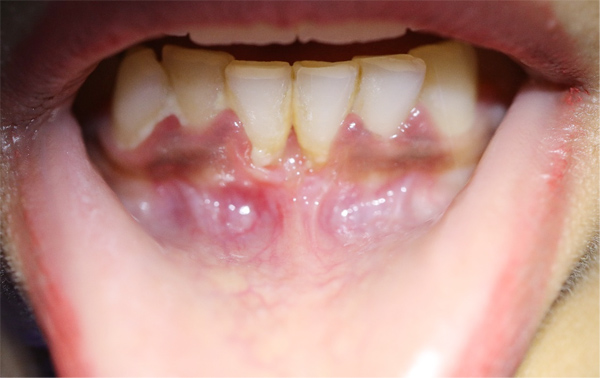
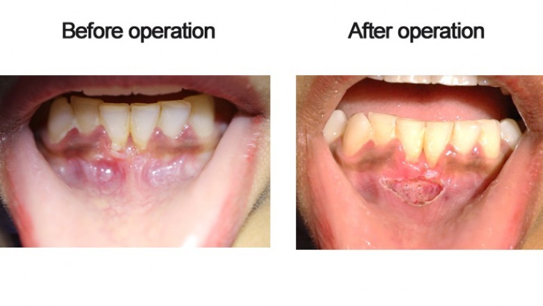
Preoperative View (Courtesy- Dr. Sana Farista)
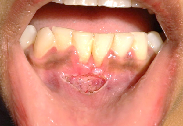
Immediate Postoperative view (Courtesy- Dr. Sana Farista)
Reference - Patil P, Kabbur KJ, Madaiah H, Satyanarayana S. Diode laser frenectomy: A case report with review of literature. J Dent Lasers 2019;13:19-22 and Patel R M, Varma S, Suragimath G, Abbayya K, Zope S A, Kale V. Comparison of labial frenectomy procedure with conventional surgical technique and diode laser. J Dent Lasers 2015;9:94-9